
A. COMPONENTS OF A CELL STRUCTURE
• CELL MEMBRANE/PLASMA MEMBRANE—Outermost layer of the cell
• PROTOPLASM –A complex material within which the cell is enclosed
• NUCLEUS—Central part of the cell
• CYTOPLASM—Outer dense less part of the protoplasm
• CYTOSOL-fluid matrix of the cytoplasm
• VACUOPLASM—The spaces within which the membranes are enclosed
B. FLUID MOSAIC MODEL
The structure of the cell membrane is called a fluid mosaic model. It consists of the composition of lipids, carbohydrates, and proteins.
-
Lipids :
Each phospholipid molecule has two portions—head and tail. The head end is called the polar end and the tail end is called the non-polar end. The head part is said to be hydrophilic because it is soluble in water and the tail part is called hydrophobic because it is insoluble in water. The dark-staining parts of the membranes are formed by the head of the molecules and the light-staining intermediate zone is made by the tails.
-
Proteins :
These are embedded in the thickness of the membranes. Some proteins occupy the entire thickness of the
membrane and bulge out of both its surface and are called transmembrane proteins. Some proteins form passive channels through which the substances can diffuse through the membrane. Some proteins act as receptors for specific hormones.
-
Carbohydrates
The carbohydrate layer is well developed on the external surface of the plasma membrane and forms the cell boundary. This layer is called glycocalyx.
C. FUNCTIONS OF THE CELL MEMBRANE
- Maintains the shape of the cell and controls the passage of all substances in or out of the cell.
- Certain receptors are present on the surface of the cell membrane that contains enzymes and hormones.
- Membrane proteins help to maintain the structural integrity of the cell.
- . Some cell membranes may show a high degree of specialization.
D. TRANSPORT OF MATERIALS ACROSS THE CELL MEMBRANE
Transport across the cell membrane can either be passive or active. Passive diffusion can either be by simple diffusion or
facilitated diffusion.
- Endocytosis is the process of actively transporting molecules into the cell.
- Phagocytosis is the process by which the cell engulfs some of the foreign particles.
- Pinocytosis is the process by which the vesicles of the cell are used for the absorption of fluids into the cell.
- Exocytosis is a form of active transport in which a cell transports molecules out of the cell.
E. CELL JUNCTIONS
- Intercellular connections between adjacent cells of animal tissues.
- These are formed by multiprotein complexes and can be recognized by electron microscope.
- There are 3 major types of cell JUNCTIONS— anchoring junctions/ adhesive junctions that bind cell together; Occluding junctions/ tight junctions; and communicating junctions/ gap junctions.
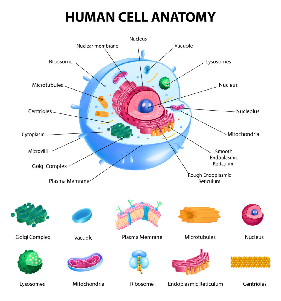
F. CELL ORGANELLES
- Generally, the cytoplasm of the cell contains various structures, referred to as organelles.
- These include the endoplasmic reticulum, ribosomes, mitochondria, Golgi complex, and vacuoles.
1. ENDOPLASMIC RETICULUM
Network of membranous tubules and it is continuous with the nuclear membrane. It is involved in lipid and protein synthesis. Membranous channels are arranged in flattened sacs called cisternae. The cytoplasm within these channels is called vacuoles and that outside these channels is called hyaloplasm or cytosol.2 types- Smooth and rough endoplasmic
reticulum.
2. RIBOSOMES
Present in Rough Endoplasmic Reticulum. Lie free in the cytoplasm. There are two types- monosomes (present singly) and polysomes ( present in groups). Each ribosome consists of proteins and RNA and is 15 nm in dm.
3. MITOCHONDRIA
Double membrane-bound organelle. This can be seen under a light microscope in stained preparations. Appears as granules or as rods. These are large in cells with a high oxidative metabolism. Bounded by a smooth outer membrane within which there is an inner membrane. The inner membrane has incomplete partitions called Cristae. The space
bounded by the inner membrane is filled by a granular material called the matrix. It is called the powerhouse of the cell. ATP and GDP are formed in mitochondria.
4. GOLGI COMPLEX
Membrane-bound organelles of the cell and is made up of a series of flattened, stalked pouches called cisternae. Within under the light microscope, the Golgi complex can be seen as a small structure of irregular shape, present near the
nucleus. The Golgi apparatus is made up of four to eight cisternae and in some single-celled organisms, it may consist of 60 cisternae. The apparatus has three regions known as cis, median, and trans.
5. LYSOSOMES
These vesicles contain enzymes that destroy the unwanted material present within a cell. These enzymes include proteases, lipases, carbohydrates, and acid phosphatase. These digest the materials within phagosomes.
6. PEROXISOMES
These are similar to lysosomes because they are membrane-bound vesicles containing organelles. Their enzymes– hydrogen peroxide is used to detoxify various substances by oxidizing them. These are prominent in cells of the liver and
renal tubules.
7. CYTOSKELETON
A network of filaments and tubules that extends throughout a cell and cytoplasm. The cytoplasm supports the cell, gives it shape, organizes, and tetheres the organelles. The elements that constitute the cytoskeleton consist of— microfilaments, microtubules, and intermediate filaments.
8. CENTRIOLES
Small structure made up of microtubules and exists as part of centrosomes. Important for the formation of various cellular structures that are made up of microtubules. Under the light microscope, the two CENTRIOLES are seen as dots embedded in a region of dense cytoplasm, called the centrosome.
9. NUCLEUS
Central,dense part of the cell and is rounded or ellipsoidal. It stores the cells hereditary material or DNA, and coordinates the cell activities.The materials making the fibres of the network in nucleus is called chromatin.Chromatin is seen in the form of irregular dark masses that are called heterochromatin. Nuclei in which there are large areas of euchromatin can be called as open face nuclei. Nuclei that are made up mainly of heterochromatin are referred to as closed face nuclei. The nucleolus is a membranous organelle within the nucleus that makes the ribosomes. Nucleoplasm is the space between the various constituents of the nucleus. Under the microscope,the nucleus is seen to be surrounded by a double layered nuclear membrane or nuclear envelope.
10. NUCLEOLI
These are spherical and contains one or more nucleoli. Stains both in hematoxylin and eosin.Have a central filamentous zone — pars filamentosa and an outer granular zone — pars granulosa and both of these are embedded in an amorphous material — pars amorpha. Parts of the chromosomes located within nucleoli constitute the pars chromosoma of nucleoli.
11. CILIA
Hair like projections from free surfaces of some epi cells. It is motile in living animals. Free part of each cilium is called the shaft and the region of attachment of the shaft to the cells surface is called the base. Each cilium is 0.25 micrometer in dm. Microtubules of cilia are bound with two proteins— dynein and nexin. Cilia present on the cells in the olfactory mucosa of the nose are called olfactory cilia.Kinociliaare are present in some parts of the internal ear.
12. FLAGELLA
Larger processes with the same functions as cilia. The best example of flagellum is the tail of the spermatozoa.
13. STAINING OF A CELL
Nuclei are stained blue,whereas the cytoplasm and extracellular matrix have pink staining.
Leave a Reply to meetwe08 Cancel reply